Biomedical Devices

Magnetic Collection of Joint-Level Osteoarthritis Biomarkers

Motivation

Diagnosis of early-stage osteoarthritis (OA), a disease stage where emerging  therapeutics have demonstrated potential to reduce and prevent OA progression in animal models, remains a significant clinical challenge. However, OA early-stage detection could lead to interventions and change in lifestyle that would reverse the chronic cascade of joint destruction found in the OA-affected joint. Clinically, OA is diagnosed through radiographs and physical exams, yet these diagnostics are relatively poor at detecting early-stage OA. A significant need exists for technologies that facilitate early-stage OA diagnosis. Therefore, direct assessment of molecular changes within an OA-affected joint would overcome these limitations.  The goal of this project is to develop a novel magnetic nanoparticle-based technique to collect OA biomarkers from synovial fluid without the need to remove fluid from the joint space.

Our preliminary studies demonstrate a proof-of-concept for magnetic harvesting; however, additional refinement is needed to accurately and repeatedly relate the amount of biomarker collected to the initial biomarker concentration in the synovial fluid.


Spatially controlled electrospun solid gradient nanofibers for guided spiral ganglion neuron culture

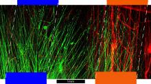
The direction of cell growth is associated with chemical, structural and/or mechanical properties of the substrate. Structurally, electrospun nanofibers provide a suitable environment for cell attachment and proliferation due to their similar physical dimension to that of the extracellular matrix. Furthermore, by modulating the topographical features of nanofibers, which include fiber diameter and orientation, cell growth and its related functions can be modified. Here, we demonstrate a solid gradient scaffold for directional growth of spiral ganglion neurons (SGNs). Spatial nanofiber alignment is controlled using a custom directional electrospinning setup. The electric field to spatially control the confinement of nanofibers was simulated with COMSOL Multiphysics simulation tool and experimentally verified. To promote neurite outgrowth and impart directionality to SGN cells, a spatial gradient of neurotrophin (NT) is introduced. By sequentially electrospinning solutions of increasing concentrations of NT in the biodegradable polymer and collecting sections of these aligned fibers along a uniaxial direction, we achieved uniform sectional nanofibers of increasing gradient concentrations. Initial tests with SGNs show improved cell adhesion and decreased morbidity to microfabricated PLGA scaffolds. Our solid gradient nanofiber membrane is versatile, obviates the need for the complex microfluidic mixer system to generate an NT gradient, is potentially implantable, and can be used in other nerve regeneration studies in peripheral nerve system and central nerve system.

Fabrication of Nanoporous membrane and its non-lithographic patterning using Electrospinning and Stamp-thru-Mold (ESTM)

The Electrospinning and Stamp-thru-mold (ESTM) technique, an integrated fabrication process which incorporates the versatility of the electrospinning process for nanofiber fabrication with
the non-lithographic patterning ability of the stamp-thru-mold process is introduced. In-situ multilayer stacking of orthogonally aligned nanofibers, ultimately resulting in a nanoporous membrane, has been demonstrated using orthogonally placed collector electrode pairs and an alternating bias scheme. The pore size of the nanoporousmembrane can be controlled by the number of layers and the deposition time of each layer. Non-lithographic patterning of the fabricated nanoporousmembrane is then performed by mechanical shearing using a pair of pre-fabricated micromolds. This patterning process is contamination free compared to other photo lithographical patterning approaches. The ability to pattern on different substrates has been tested with and without oxygen plasma surface treatment. In vitro tests of ESTM poly-lactic-coglycolic acid (PLGA) nanofibers verify the biocompatibility of this process. Simulation by the COMSOL Multiphysics tool has been conducted for the analysis of electrospun nanofiber alignment.